HUMAN BRAIN ANATOMY
The brain is composed of several parts that control what you think; how you feel; where you move; what you see, hear, and taste; and many other functions. The cerebrun, cerebellum, and diencephalon are some of the major sections of the brain.
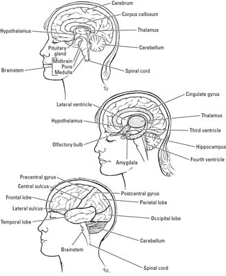
The cerebrum
The cerebrum is made up of the left and right cerebral hemispheres, separated by the falx cerebri. Each hemisphere has five lobes: the frontal lobe in the front (of course!), the temporal and parietal lobes on the sides, the occipital lobe in the back, and the insular lobe located between the temporal lobe and the frontal lobe. The cerebral cortex is the outer layer of the cerebrum. The surface of the cerebral cortex isn’t smooth; it has folds, grooves, and clefts. The folds are called gyri, the grooves are sulci (singular: sulcus), and the clefts are called fissures. These features increase the surface area of the brain while still allowing into to fit into its bony vault.
The cerebral hemispheres are connected by the corpus callosum, a band of nerve fibers that allows each side to communicate with the other. The cingulate gyrus is located superior to the corpus callosum. It helps coordinate emotions. The hippocampus and the amygdala are located in the temporal lobe and are important for memory.
The cerebrum has many different higher functions. It’s involved in controlling cognitive functions; shaping your personality, feelings, and perceptions; and handling motor functions and sensory interpretation.
The diencephalon
The diencephalon includes the epithalamus (the posterior part), thalamus (the middle part), and hypothalamus (the most inferior part). The diencephalon serves as a relay station, interconnecting different parts of the nervous system, and controls many autonomic nervous functions.
The cerebellum
The cerebellum is the portion of the brain lying beneath the tentorium cerebelli in the posterior part of the cranium. It’s made up of two hemispheres connected by the narrow wormlike part of the cerebellum called the vermis. The cerebellum controls balance, coordinates movement, and maintains muscle tone.
The brainstem
The brainstem includes three parts: the midbrain is the most superior part, the pons is in the middle, and the medulla oblongata (medulla) is the most inferior portion and connects to the spinal cord. The brainstem controls your levels of alertness, arousal, respiratory rate, blood pressure, digestion, heart rate, and other autonomic functions.
Cerebrospinal fluid protects the brain from damage by cushioning it during a blow to the head and by helping hold up the brain, which takes the pressure off the base of brain. The fluid is produced by the choroid plexus, which is located in four ventricles in the brain. Two lateral ventricles extend into the cerebral hemispheres. They open into the third ventricle through the interventricular foramina. The fourth ventricle is located in the brain stem and is connected to the third ventricle by cerebral aqueduct, or aqueduct of Sylvius. The fourth ventricle is connected to the central canal. The median aperture (also called the aperture of Magendie) and the two lateral apertures (or apertures of Luschka) connect the fourth ventricle to the subarachnoid space.
The cranial cavity contains two glands: the pituitary gland and the pineal gland. They’re tiny little things, but they’re so important, especially the pituitary gland. It’s so significant that it’s referred to as the master gland.
Pituitary gland: This small gland is about the size of a pea. It’s located in the sella turcica of the sphenoid bone. It is connected to the hypothalamus by a stalk called the infundibulum. It’s called the master gland because it secretes several hormones that influence the activity of many other endocrine glands that regulate a variety of body functions, including blood pressure, childhood growth, urine production, testosterone production in males, and estrogen production in females. It has two parts: the anterior portion, the adenohypophysis, and the posterior portion, called the neurohypophysis.
Pineal gland: Also called the epiphysis, this gland is a small pine-cone-shaped body located at the posterior end of the roof of the third ventricle. It synthesizes melatonin, a hormone that regulates sleep and circadian rhythm based on the body’s exposure to light. A problem with the pineal gland can lead to a lot of sleepless nights.


0 Comments